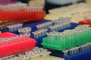
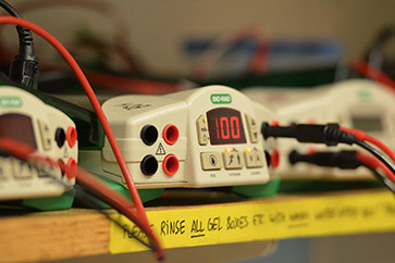
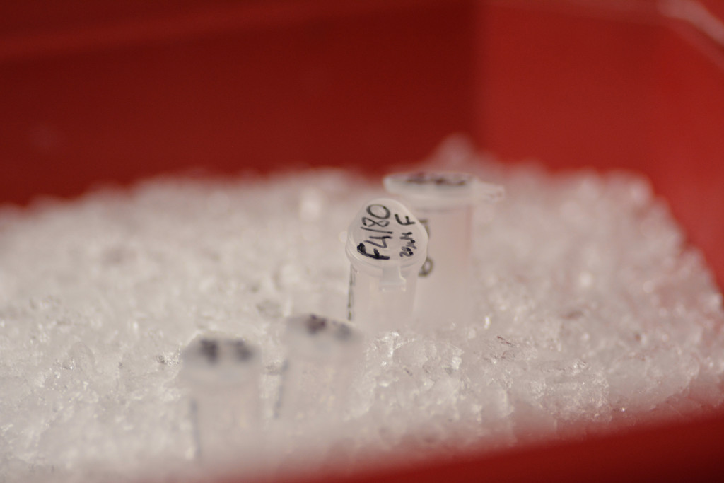
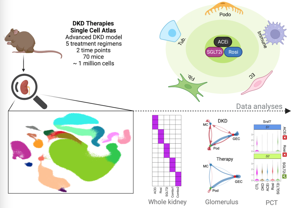
single cell multiomics
We use single cell technologies to drive precision medicine
kidney organoids
We generate kidney organoids to model development & disease.
regenerative med
We develop treatments for human kidney diseases.


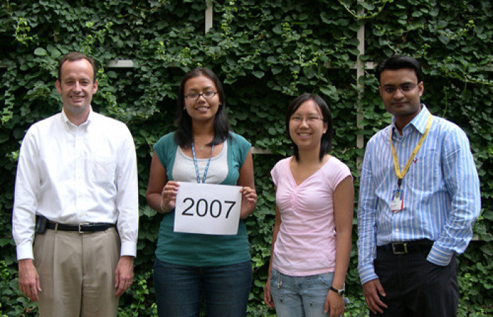
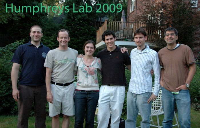
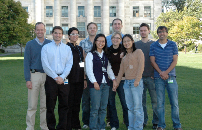
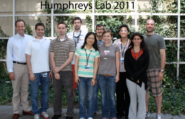
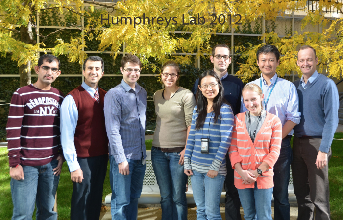
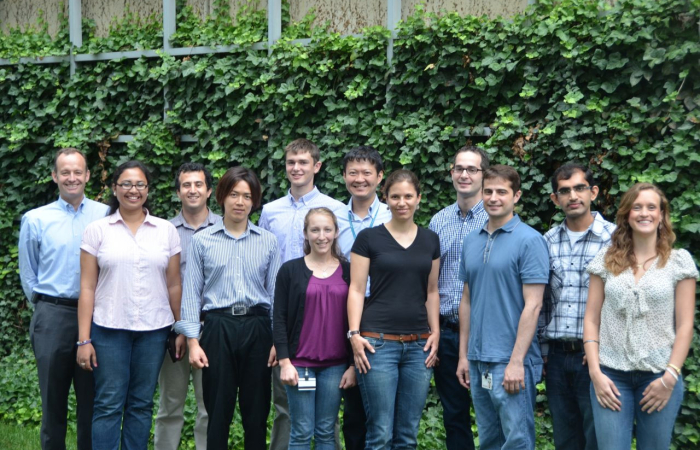
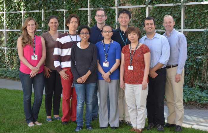
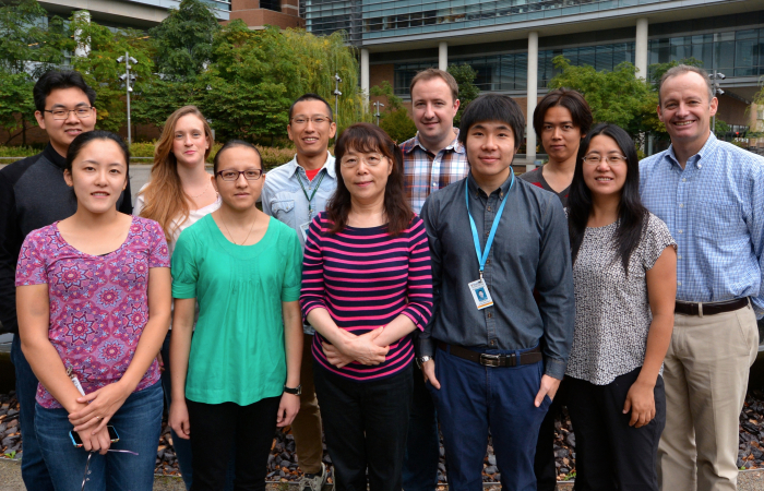
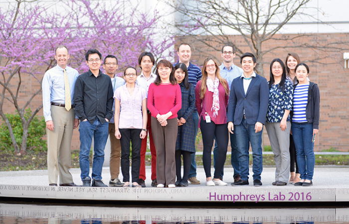
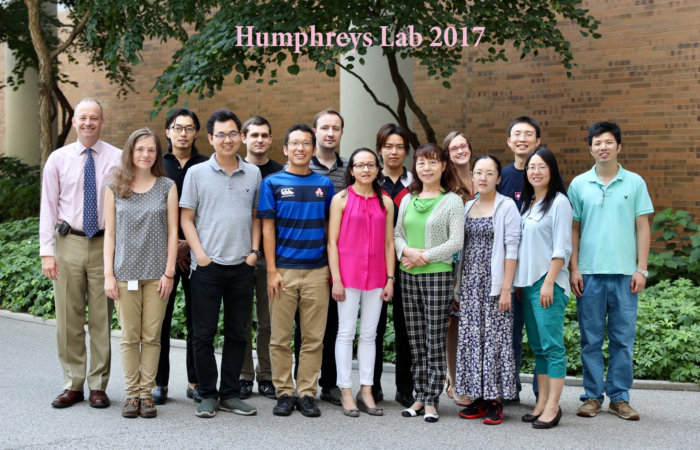
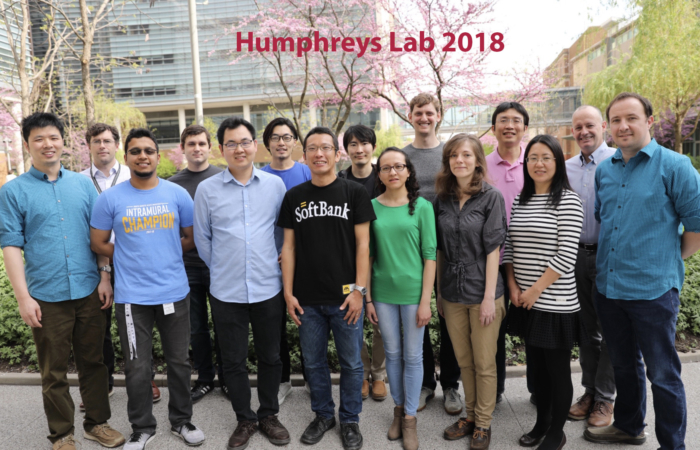
Watch the lab grow!